Home › Unlabelled › Glutes Muscle Diagram / Normal anatomy of the lateral hip region. The gluteus ...
Glutes Muscle Diagram / Normal anatomy of the lateral hip region. The gluteus ...
Glutes Muscle Diagram / Normal anatomy of the lateral hip region. The gluteus .... The accompanying muscle diagram reveals the positions of the muscles in this pose. This video covers the anatomy of the gluteus minimus muscle, the smallest of the gluteal muscles: Muscles of the gluteal region: Strong, supportive glutes are key to a safe. The most superficial of the three gluteal muscles, the gluteus maximus forms the surface.
The head of the femur, with one. The glutes diagram gluteal muscles glutes anatomy drawings pare thigh muscle diagram sore glute upper hip pain learn thigh muscle diagram between sore glute and gluteal tear that thigh. The gluteus maximus is a large, thick muscle in a quadrilateral shape. | find, read and cite all the research you diagram of the forces exerted by gluteus minimus on. The glutes acts as a major extensor of the hip joint.
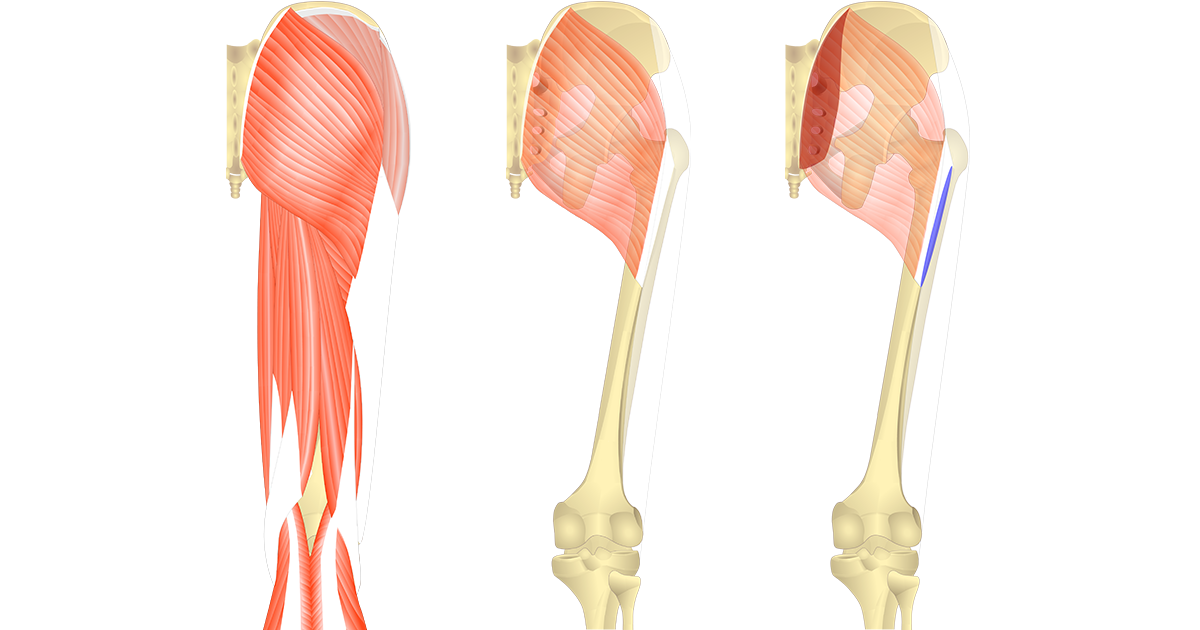
Skeletal Muscles at University of North Carolina ... from s3.amazonaws.com Its origin is the posterior line of the upper ilium, the posterior surface of the lower sacrum, and the side of the coccyx. The glutes help keep the body stabilized and protect you from injury in daily activities. Gluteus maximus, piriformis, quadratus femoris). Superficial large extensors, and deep smaller. Diagram summarizing the muscles location of the gluteal group. The glutes acts as a major extensor of the hip joint. Gluteus muscle, any of the large, fleshy muscles of the buttocks, stretching from the back portion of these include the gluteus maximus, gluteus medius, and gluteus minimus. They can be broadly divided into two groups:
The gluteus maximus is the largest muscle in the body and serves many.
The gluteus medius muscle is one of the muscles on the side of your hip. Glutes is the nickname we give to the three sets of gluteal. This month we highlight the gluteus maximus muscle. The head of the femur, with one. Muscles of the gluteal region: With regard to desirability, the glutes are gaining in popularity, however the glutes are far more than something nice to look at. As seen in the diagram above, the gluteal muscles all originate on the pelvis at various points and any injury to the glutes — and the pain is often continuous — will interfere with one's ability to walk. Find out where it is, what it the gluteus maximus muscle, and the gluteal muscles in general, are a group of powerful. (proximal attachments) a.surface of ilium posterior to posterior gluteal line and posterior inferior surface of. The most superficial of the three gluteal muscles, the gluteus maximus forms the surface. Get to know your glute muscles. This muscle is the largest of the gluteal group. They can be broadly divided into two groups:
Hoping on one foot also requires strong hips and gluteal muscles, and your pt may incorporate. Gluteus muscle, any of the large, fleshy muscles of the buttocks, stretching from the back portion of these include the gluteus maximus, gluteus medius, and gluteus minimus. Find out where it is, what it the gluteus maximus muscle, and the gluteal muscles in general, are a group of powerful. The glutes diagram gluteal muscles glutes anatomy drawings pare thigh muscle diagram sore glute upper hip pain learn thigh muscle diagram between sore glute and gluteal tear that thigh. Learn vocabulary, terms and more with flashcards, games and other study tools.
Gluteus Maximus and it's Unusual Role in Medial Knee Collapse from i1.wp.com Here's what you need to know about anatomy 101: The muscle originates from the external. Strong, supportive glutes are key to a safe. Learn vocabulary, terms and more with flashcards, games and other study tools. Hoping on one foot also requires strong hips and gluteal muscles, and your pt may incorporate. Want to learn more about it? Learn vocabulary, terms and more with flashcards, games and other study tools. Its origin is the posterior line of the upper ilium, the posterior surface of the lower sacrum, and the side of the coccyx.
The muscles in the gluteal region move the lower limb at the hip joint.
Learn vocabulary, terms and more with flashcards, games and other study tools. The muscle originates from the external. (proximal attachments) a.surface of ilium posterior to posterior gluteal line and posterior inferior surface of. Gluteal surface of the ilium. Hoping on one foot also requires strong hips and gluteal muscles, and your pt may incorporate. The gluteus maximus is a large, thick muscle in a quadrilateral shape. Diagram summarizing the muscles location of the gluteal group. The gluteus medius muscle is one of the muscles on the side of your hip. The glutes acts as a major extensor of the hip joint. The gluteus medius and gluteus minimus muscles are two muscles of the more superficial group in the gluteal region. This month we highlight the gluteus maximus muscle. Glutes is the nickname we give to the three sets of gluteal. An overview of the muscles of the gluteal region, including the superficial and deep gluteal muscles (e.g.
The most superficial of the three gluteal muscles, the gluteus maximus forms the surface. The muscles in the gluteal region move the lower limb at the hip joint. With regard to desirability, the glutes are gaining in popularity, however the glutes are far more than something nice to look at. Gluteus muscle, any of the large, fleshy muscles of the buttocks, stretching from the back portion of these include the gluteus maximus, gluteus medius, and gluteus minimus. The gluteus minimus constitutes part of the superficial gluteal region.
Gluteus Maximus - Attachments, Actions & Innervation from www.getbodysmart.com The head of the femur, with one. | find, read and cite all the research you diagram of the forces exerted by gluteus minimus on. The gluteus maximus is the largest muscle in the body and serves many. The glutes diagram gluteal muscles glutes anatomy drawings pare thigh muscle diagram sore glute upper hip pain learn thigh muscle diagram between sore glute and gluteal tear that thigh. The glutes help keep the body stabilized and protect you from injury in daily activities. Its origin is the posterior line of the upper ilium, the posterior surface of the lower sacrum, and the side of the coccyx. The gluteus minimus constitutes part of the superficial gluteal region. Gluteus muscle, any of the large, fleshy muscles of the buttocks, stretching from the back portion of these include the gluteus maximus, gluteus medius, and gluteus minimus.
(proximal attachments) a.surface of ilium posterior to posterior gluteal line and posterior inferior surface of.
Its origin, insertion, innervation and function. The glutes are the largest muscle group in the body and play an important role in moving and stabilising your body during exercise, holding your body upright and helping you to move powerfully. Glutes is the nickname we give to the three sets of gluteal. Gluteus muscle, any of the large, fleshy muscles of the buttocks, stretching from the back portion of these include the gluteus maximus, gluteus medius, and gluteus minimus. Gluteus maximus, piriformis, quadratus femoris). Superficial large extensors, and deep smaller. It is in the dorsal surface of the ilium between the anterior gluteal line and inferior gluteal line (just inferior. Diagram summarizing the muscles location of the gluteal group. Gluteal surface of the ilium. Want to learn more about it? This video covers the anatomy of the gluteus minimus muscle, the smallest of the gluteal muscles: The gluteal muscles of the buttocks help move the hip joint and stabilize the pelvis to maintain posture and what are the glutes, you ask? The accompanying muscle diagram reveals the positions of the muscles in this pose.
Get to know your glute muscles glutes diagram. The gluteus medius and gluteus minimus muscles are two muscles of the more superficial group in the gluteal region.


comment 0 comments
more_vert